West Bengal Nursing Council

Indian Nursing Council, New Delhi

Director of Medical Education (DME)

Pharmacy Council of India, New Delhi

West Bengal University of Health Sciences

Recognition of College Section 2 (f) of the UGC ACT 1956

QS I-Gauge E-LEAD Certified for Excellence in Academic Digitisation

MAKAUT, Formerly WBUT, West Bengal

All technical courses are approved by AICTE, Ministry of Education, Govt. of India
Programme & School Offered
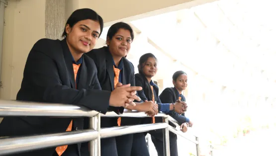
Where Curiosity Meets Academic Excellence


Browse By Schools
Placements
Every student tells a story of success
Nurtured through innovation, strong values, and a learning environment that empowers future leaders.


Swagata Nayak
placed at Rinex


Securing a placement through NSHM was a milestone moment in my academic journey
Simran Prasad
placed at TCS


Securing a placement through NSHM was a milestone moment in my academic journey
Utsav Narayan Deb
placed at Auberge de Cassagne & Spa, France


Manmeet Kaur
placed at Adidas


Shreyashi Dewasi
placed at Aig Hospital


Arhan Khan
placed at Medanta


Anushka Ghosh
placed at Max Healthcare


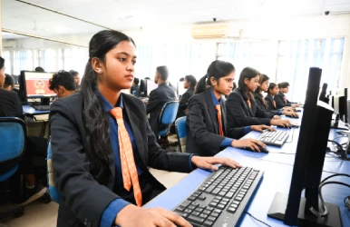
Facilities
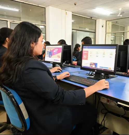
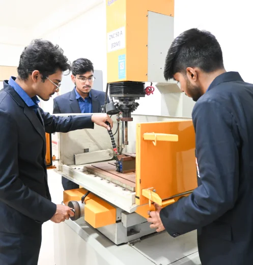
Smart Spaces for Smarter Minds
We are one of the top colleges in Durgapur and our advanced laboratories provide students with practical exposure to cutting-edge tools, technologies, and real-world industry applications.

Modern Campus Infrastructure
As one of the best private colleges in Durgapur, we offer state-of-the-art classrooms, computer centers, laboratories, a well-stocked library, dedicated research labs, and specialized spaces


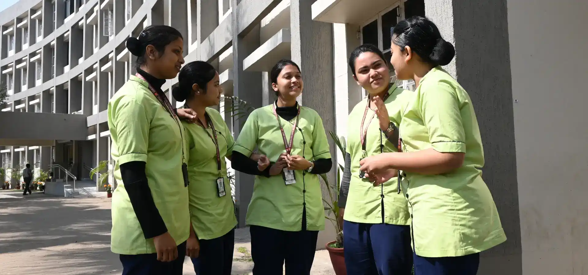
About Us
Shaping Excellence with Modern Learning Spaces
The NSHM Durgapur campus offers a comprehensive academic environment with state-of-the-art infrastructure and facilities aligned with global standards. We have also emerged as one of the best private colleges in Kolkata.


Indian Nursing Council, New Delhi

Director of Medical Education (DME)

Pharmacy Council of India, New Delhi

West Bengal University of Health Sciences

Recognition of College Section 2 (f) of the UGC ACT 1956

QS I-Gauge E-LEAD Certified for Excellence in Academic Digitisation

MAKAUT, Formerly WBUT, West Bengal

All technical courses are approved by AICTE, Ministry of Education, Govt. of India
Years of Academic Excellence
Strong Alumni Network
Industry Associates
Success Stories
Explore student and alumni success stories of dedication, growth, achievement.


Request a Call